Bone structure of knee joint-femur
 Apr. 15, 2020
Bone structure of knee joint-femur
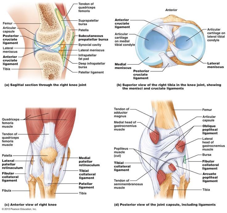
The femur, tibia and patella are involved in the formation of the knee joint. The above three bones correspond to each other and form three relatively independent medial compartments of the knee joint, the lateral ventricle and the patellofemoral joint compartment.
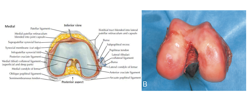
The distal femur is the attachment site of many ligaments and tendons, and the anatomical shape is more complex (see Fig.1). In terms of shape and size, the internal and external condyle of the femur are not symmetrical.


The medial femoral condyle was larger with the same curvature on the sagittal plane, the lateral femoral condyle was smaller, the curvature of the lateral femoral condyle was gradually increasing from the forward to the posterior, and the axis of the lateral condyle of the femur was slightly shorter than that of the medial condyle. The angle between lateral femoral condyle axis and sagittal plane is smaller than that between medial femoral condyle axis and sagittal plane, and the angle between the latter and sagittal plane is 22°. The lateral femoral condyle is slightly larger than the medial condyle if the intercondylar fossa of the femur is the midpoint.
The front of the femoral condyle is separated by a groove, the femoral trochlear (see Fig.2). The deepest part of the femoral trochlear is called the trochlear groove. The trochlear groove is slightly more lateral than the median plane between the internal and external condyles. Accurate reconstruction of the above anatomical relationship is essential to restore normal biomechanics of patellofemoral joint.


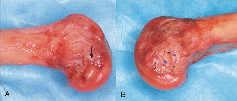
The external epicondyle of the femur is smaller but more prominent. It is the starting point of lateral collateral ligament or fibular collateral ligament. There are more ascending adductor tubercle on the medial femoral condyle, and the great adductor muscle is here. The medial epicondyle is located in the far anterior part of the adductor tubercle, which is a "C" shaped ridge uplift (see Fig.3)

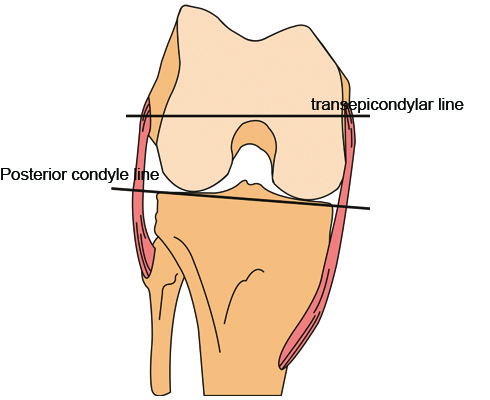
The central depression of the medial condyle is in the form of a shallow groove , the medial collateral ligament is the ridge - like ridge of the sulcus rather than the outer edge . The medial and external condylar line of the femur refers to the axis ( see Fig.4 ) at the highest point of the medial condylar groove and the superior point of the outer superior condyle . The line is an important reference axis during total knee arthroplasty.

For normal knee joint, if the posterior condyle is the reference osteotomy, the average external rotation of the line of the internal and external epicondyle of the femur is 3.5°for the male and 1°for the female. For the patients with osteoarthritis and genu valgus, the external rotation of the line of the medial and lateral epicondyle of the femur can reach more than 10°.


