Management of Combined Deficits With Structural Grafting
 Nov. 09, 2020
Management Of Combined Deficits With Structural Grafting
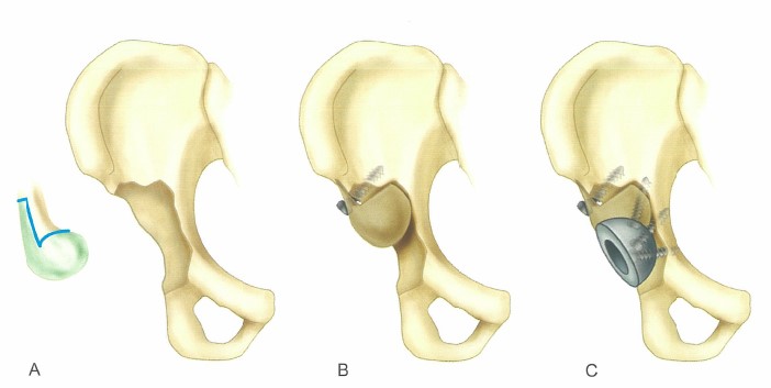
■ Select a distal femoral allograft of appropriate size to fill the defect. Shape the condylar portion of the graft with reamers so that the size of the graft is approximately 2 mm larger than the defect. Fashion the graft in the shape of an inverted 7.
■ Make a diaphyseal cut in the coronal plane, leaving the anterior cortex thick enough for screw fixation.
■ Make an oblique cut through the posterior cortex, exiting just proximal to the posterior condyles (Fig. 3-161A).
■ Place the graft over the defect in the superior portion of the acetabulum, and gently impact it into place with the cut portions of the graft buttressed against the superior portion of the acetabulum and ilium.
■ Secure the graft to the ilium with multiple screws or a plate. Place the screws in a staggered fashion, and tap the drill holes to avoid fracturing the graft (Fig. 3-161B).
■ Ream the graft and remaining portions of the acetabulumwith standard acetabular reamers to a size that leaves the anterior and posterior columns intact, while maximizing contact for bone ingrowth for the revision prosthesis (Fig. 3-161C).
■ Impact the acetabular component in the appropriate position and fix it to the host bone with multiple screws.


FIGURE 3-161 Paprosky " 7 " graft for segmental acetabular deficiency.
A, Distal femoral allograft is shaped to resemble numeral 7.
B, Graft is shaped to fit acetabular deficiency and fixed as shown , with several screws placed above acetabulum through remaining cortical portion of graft.
C, Graft is reamed, and revision component is implanted.
When segmental and cavitary deficits occur simultaneously, the segmental defect is reconstructed first to restore the rim. Any remaining cavitary deficits are filled with particulate cancellous graft.
Note: this article comes from CAMPBELL’S OPERATIVE ORTHOPAEDICS by S. Terry Canale James H. Beaty.


