Removal of femoral cement
 Jun. 01, 2020
REMOVAL OF FEMORAL CEMENT (TECHNIQUE 3-15)
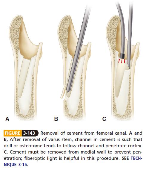
■ To remove the proximal cement, clear soft tissue andoverhanging cement away from the proximal femur toexpose the proximal edge of the bone-cement interface.Remove any cement and overhanging bone from thegreater trochanter first to allow unobstructed passage ofinstruments directly down the canal. This is especiallyimportant if the previous stem was placed in varus. If thetrochanter blocks passage of instruments directly downthe canal, the instruments are likely to exit through aperforation created in the posterolateral cortex (Fig.3-143). Osteotomize the greater trochanter if necessary.
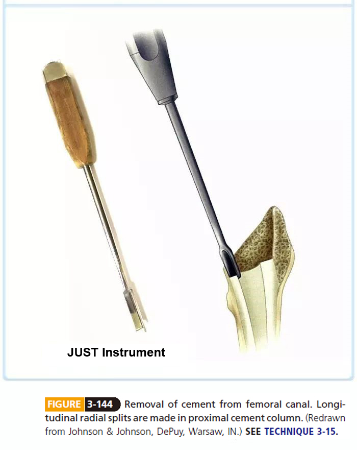
■ Make numerous longitudinal radial splits in the proximalcement column (Fig. 3-144).
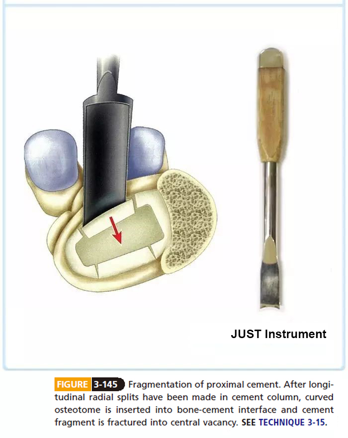
■ Insert osteotomes into the bone-cement interface, andfracture the fragments of cement into the open centralarea (Fig. 3-145). Do not attempt to wedge instrumentsinto the bone-cement interface before making the radialsplits, or a femoral fracture is likely to occur.
■ Removal of cement from around the middle and distalsegments of the stem is more difficult, although it stillcan be accomplished with hand tools. Adequate lighting,irrigation, and suction become more important to showthe bone-cement interface. The available space centrallynarrows because of the taper of the stem. Narrow, angledcement osteotomes are often required.
■ Small cement fragments may occlude the central space.Remove them with a pituitary rongeur and curets.
■ Review the preoperative radiographs to determine thickand thin areas of cement to be removed and to determineany deviation of the tip of the stem that may direct instrumentsout through the cortex rather than centrally in thecanal. Also take into account the normal anterolateralbow of the femur. Sometimes this segment of the cementmantle can be removed en masse by passing a largethreaded tap into the defect left by the stem and extractingit with a slap hammer.
■ Removal of the distal cement mass usually is the mostdifficult. If the plug of cement is thin and not tightlyadherent to the cortex, it occasionally can be driven distally in the canal and left. This should not be done inthe presence of infection.
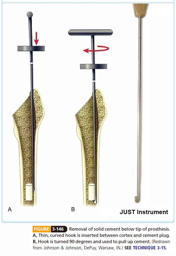
■ If the plug is thin and does not fill the canal completely,it often can be extracted with a hook. Pass the hookbetween the cortex and cement through any apparentgaps on the preoperative radiographs (Fig. 3-146A). Turnthe hook 90 degrees to engage the cement, and gentlytap the hook with a mallet to withdraw the cement plug(Fig. 3-146B).
■ Fragmentation of solid distal cement with cruciate-shapedosteotomes can be attempted, but this technique isslow and unrealistic if the distal cement extends morethan 1 cm.
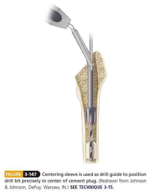
■ If the cement plug fills the canal and is firmly fixed, perforateit with a drill and use reamers to enlarge the hole.Carefully drill the initial hole in the center of the distalcement plug, and align the drill with the femoral canal.Use a centering sleeve to position the drill bit precisely (Fig. 3-147). Enlarge the hole with masonry drills speciallydesigned for this purpose. After a hole of sufficient sizehas been made, remove the remaining cement adherentto the cortex with a reverse cutting hook. Alternatively,insert a tap into the hole and extract the cement plugwith a mallet.
Note: this article comes from CAMPBELL’S OPERATIVE ORTHOPAEDICS by S. Terry Canale James H. Beaty.


